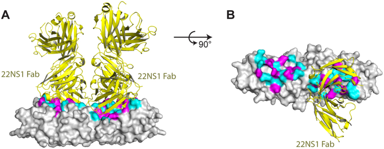
Figure 5. The epitope for WNV protective antibody 22NS1.
(A,B) The structures of 22NS1-bound WNV NS1 (pdb ID 4OII)24 and ZIKV NS1 are superimposed on their β-ladder dimers. ZIKV NS1 is represented in grey surface, and the 22NS1 is represented in yellow cartoon. The variable residues on the epitope are magenta, and the identical residues are cyan. For clarity, one 22NS1 is omitted in panel b.

