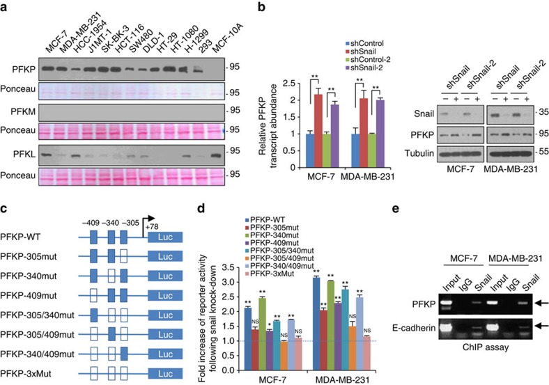
Figure 3. PFKP is a major isoform of PFK-1 in cancer cells and a target of Snail repressor.
(a) PFK-1 immunoblotting of cell lysates from various human cancer cells. In total, 5 μg of each cell lysate for PFKP and 30 μg of cell lysate for PFKM or PFKL were subjected to immunoblot analysis. The films were developed at the same exposures to compare relative protein abundance of PFK-1 isoforms, and protein loading of each blot was validated with Ponceau stain. The PFK-1 antibody validation using positive control of platelet, muscle and liver tissue are shown in Supplementary Fig. 10c. Cell lines used in this study was authenticated by short tandem repeat profiling as shown Supplementary Fig. 11. (b) Relative transcript (left) and protein (right) abundance of PFKP following knockdown of Snail (shSnail) in breast cancer cells. A total of 20 μg and 5 μg of cell lysates were used to detect endogenous Snail and PFKP, respectively. Representative blots are shown from at least two independent experiments (a,b). (c) Schematic diagram showing positions of potential Snail-binding canonical E-boxes on the PFKP proximal promoter reporter constructs. Arrow, transcription start site; empty boxes denote E-box mutant. (d) Fold increase of reporter activities in combination with wild type or mutated PFKP promoter following Snail shRNA compared with each control shRNA in breast cancer cells. *P<0.05; **P<0.01 compared with the control. (e) Chip-enriched DNA was determined by RT-PCR using specific primers complementary to the promoter regions containing E-box of PFKP (upper) and E-cadherin (lower). Statistical significances (b,d) compared with control were denoted as *P<0.05; **P<0.01 by a two-tailed Student's t-test. Unprocessed original scans of blots are shown in Supplementary Fig. 12.

