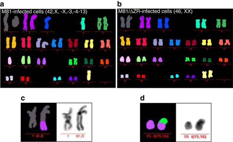
Figure 3. B cells transformed by wild-type EBV display a higher CIN rate 4 weeks post infection.
Example of a M-FISH karyotype showing mitoses from a pair of transformed cell lines infected with wild-type EBV (a), or with a replication cell-deficient mutant (b). (c,d) Two translocations are shown, found in two other cell lines transformed by wild-type EBV.

