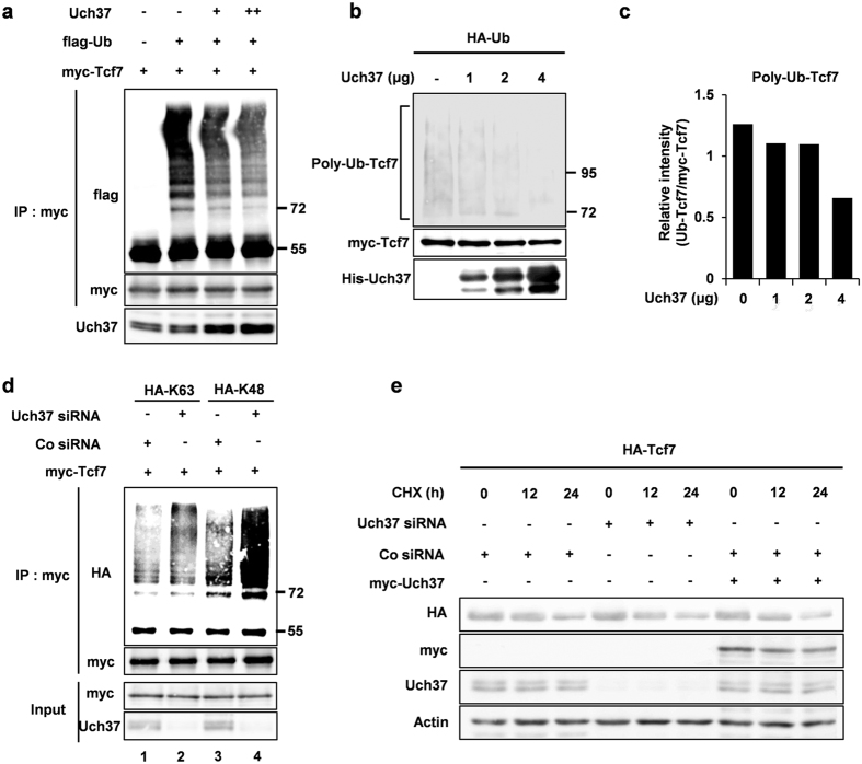
Figure 3. Uch37 deubiquitinates Tcf7 protein, but is not involved in protein stabilization.
(a) In vivo ubiquitination assay in HEK293FT cells. Cells were transfected with indicated plasmids (1 μg myc-Tcf7; 3 μg flag-Ub; 2 μg and 4 μg Uch37). After 48 h, samples were prepared as described in materials and methods and then precipitated with anti-myc antibody. (b) In vitro ubiquitination assay. HEK293FT cells were co-transfected with both myc-Tcf7 and HA-Ub plasmids to express polyubiquitinated Tcf7. After 48 h, cell lysates were immunoprecipitated using anti-myc antibody to prepare Polyubiquitinated Tcf7. Precipitated polyubiqutinated Tcf7 was then incubated with indicated amount of His-Uch37 protein. (c) Signal intensity of polyubiquitinated Tcf7 in b was quantified using image J software. (d) In vivo ubiquitination assay in HEK293FT cells. Cells were transfected with indicated plasmids and siRNAs. After 72 h, total proteins were precipitated with anti-myc antibody. Cell lysis and detailed procedures are described in materials and methods. In HA-K63, all lysines except Lys-63 were mutated to arginines. In HA-K48, all lysines except Lys-48 were mutated to arginines. (e) Pulse-chase test using HEK293FT cells. 48 hours after the transfection as indicated, 100 μg/ml cycloheximide (CHX, sigma) was treated for 0, 12, and 24 h. myc-Tcf7, Uch37, and HA-Uch37 were monitored by western blot analysis, and Actin levels were used as a loading control. Full images of all Fig. 2 are presented in Supplementary information (Figs S10 and 11).

