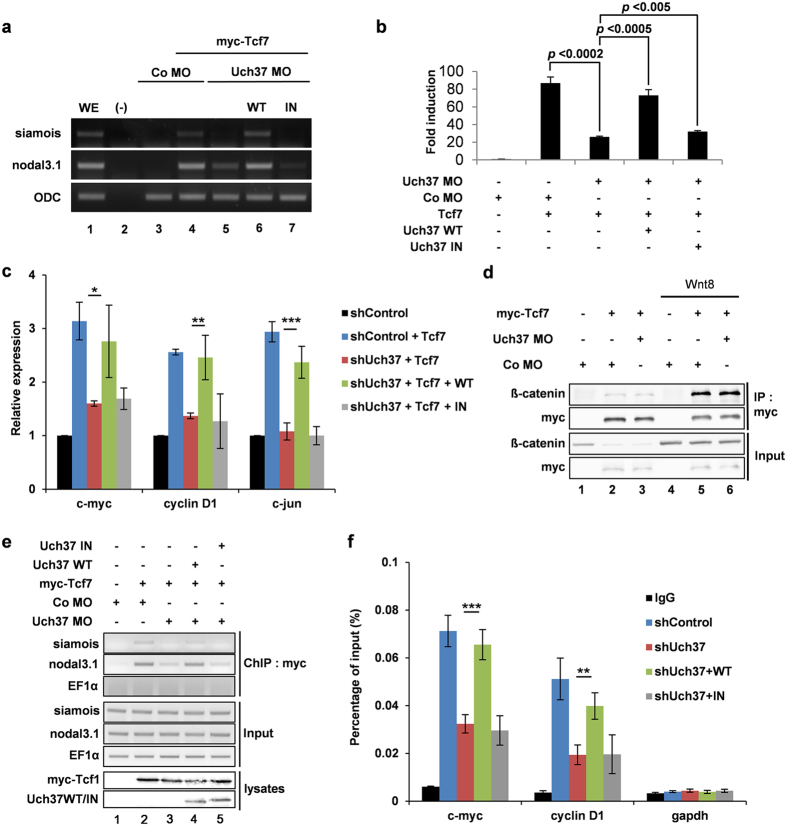
Figure 4. Deubiquitinating activity of Uch37 is required for Tcf7-mediated gene transcription by mediating DNA-binding of Tcf7.
(a) Expression levels of Wnt target genes (siamois and nodal3.1) were examined by RT-PCR analysis using Xenopus animal cap tissues. Embryos were animally injected at four-cell stage. Animal caps were isolated at stage 9, grown to stage 11. Injected reagents are as follows, 25 pg myc-Tcf7 mRNA; 20 ng Co MO; 20 ng Uch37 MO; 1 ng wild type Uch37 mRNA (WT); 1 ng catalytically inactive Uch37 mRNA (IN). Full images are presented in Supplementary information (Fig. S12). (b) TOPflash assay using whole embryos (stage 10.5, 10 embryos). Embryos were animally injected at four-cell stage. 150 pg TOPflash reporter; 50 pg pRL-TK; 40 ng Co MO; 40 ng Uch37 MO; 50 pg Tcf7 mRNA; 1 ng wild type of Uch37 mRNA (WT); 1 ng catalytically inactive Uch37 mRNA (IN). (c) qPCR analysis for the expression of Wnt-target genes (c-myc, cyclinD1 and c-jun) in stable HepG2 cells. Tcf7 was transiently transfected alone or co-transfected with wild type of Uch37 or catalytically inactive Uch37. Error bars indicate standard deviations of triplicate. *p < 0.05; **p < 0.02; ***p < 0.003 (two-tailed Student’s t test). (d) Co-IP assay using Xenopus embryos (stage 11). Two-cell stage embryos were animally injected with indicated reagents (1 ng myc-Tcf7 mRNA; 20 pg Wnt8 mRNA; 40 ng Co MO; 40 ng Uch37 MO). Total proteins were precipitated with anti-myc antibody. Active state of Wnt signalling is indicated with enhanced β-catenin level in input panel. Full images are presented in Supplementary information (Fig. S11). (e) ChIP assay using Xenopus embryos (stage 11). 70 embryos were injected at two-cell stage as indicated (25 pg myc-Tcf7 mRNA; 1 ng wild type of Uch37 (WT); 1 ng catalytically inactive Uch37 (IN); 40 ng Co MO; 40 ng Uch37 MO). Lysates were precipitated with anti-myc antibody. Precipitated Wnt target DNAs were analysed by PCR. EF1α was used as a control for specificity. Full images are presented in Supplementary information (Fig. S11). (f) ChIP assay using stable HepG2 cells. Cells were transfected with either wild type of Uch37 (WT) or catalytically inactive Uch37 (IN). Lysates were precipitated with normal rabbit IgG or anti-Tcf7 antibody. DNA-binding of Tcf7 was assessed by qPCR. gapdh was used as a negative control. **p < 0.02; ***p < 0.003 (two-tailed Student’s t test).

