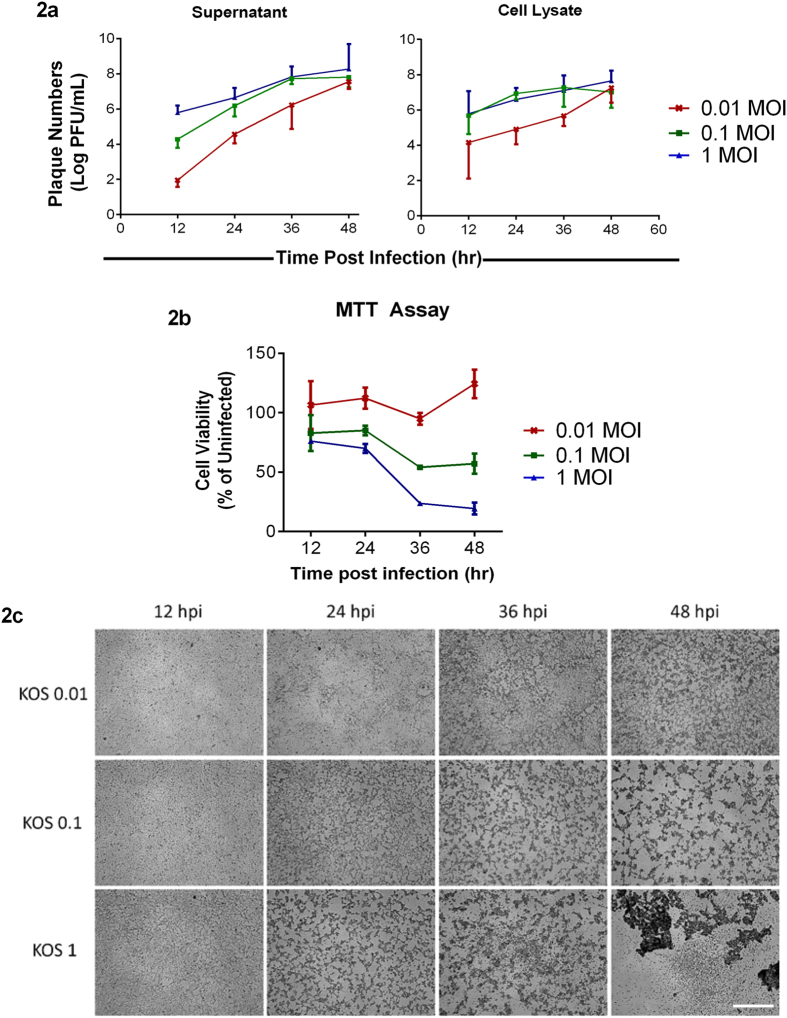
Figure 2. Initial titers determine the amount of virus released and cell viability.
(a) Supernatant and cell lysates from HCE cells infected at different MOIs were collected at the indicated times and plaque assay was performed on Vero cells as mentioned under Materials and Methods. Significance was determined by Two-way ANOVA using Tukey’s correction. Supernatant: 12 hpi: 0.01 vs 0.1, **P < 0.01; 0.01 vs 1.0, ****P < 0.0001; 0.1 vs 1.0, *P < 0.05. 24 hpi: 0.01 vs 0.1, *P < 0.05; 0.01 vs 1.0, **P < 0.01; 0.1 vs 1.0, ns. 36 hpi: 0.01 vs 0.1, *P < 0.05; 0.01 vs 1.0, *P < 0.05; 0.1 vs 1.0, ns. 48 hpi: 0.01 vs 0.1, ns; 0.01 vs 1.0, ns; 0.1 vs 1.0, ns. Cells: 12 hpi: 0.01 vs 0.1, ns; 0.01 vs 1.0, ns; 0.1 vs 1.0, ns. 24 hpi: 0.01 vs 0.1, ns; 0.01 vs 1.0, ns; 0.1 vs 1.0, ns. 36 hpi: 0.01 vs 0.1, ns; 0.01 vs 1.0, ns; 0.1 vs 1.0, ns. 48 hpi: 0.01 vs 0.1, ns; 0.01 vs 1.0, ns; 0.1 vs 1.0, ns. (b) A standard MTT assay was performed on infected HCE cells at the indicated time points. Color was read at 560 nm. Two-way ANOVA with Tukey’s correction was performed to determine significance. 12 hpi: 0.01 vs 0.1, **P < 0.01; 0.01 vs 1.0, ***P < 0.001; 0.1 vs 1.0, ns. 24 hpi: 0.01 vs 0.1, ***P < 0.001; 0.01 vs 1.0, ****P < 0.0001; 0.1 vs 1.0, ns. 36 hpi: 0.01 vs 0.1, ****P < 0.0001; 0.01 vs 1.0, ****P < 0.0001; 0.1 vs 1.0, ***P < 0.001. 48 hpi: 0.01 vs 0.1, ****P < 0.0001; 0.01 vs 1.0, ****P < 0.0001; 0.1 vs 1.0, ****P < 0.0001 (c) Representative bright field images of HCE cells at indicated times post-infection. Scale bar for all images: 500 μm.

