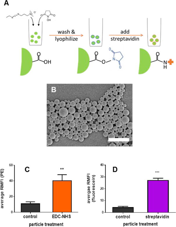
Figure 1. Particle functionalization and characterization.

(A): Schematic of preparation of streptavidin-coated particles. PLGA particles were formed using a double emulsion solvent evaporation method with simultaneous activation of terminal carboxyl groups as described in the methods section. Surface activated particles were lyophilized and coated with streptavidin immediately prior to use. (B) Representative SEM image of PLGA particles, scale bar = 2 microns. (C) EDC/NHS-activated and non-activated (control) particles incubated with streptavidin-PE. (D) EDC/NHS-activated particles treated or untreated (control) with streptavidin and then incubated with biotinylated fluorescein. When applicable, error bars = SD. ** p < 0.01, *** p < 0.001, n = 3.
