
Figure 4. Cell-particle hybrid uptake by BMDC in vitro.
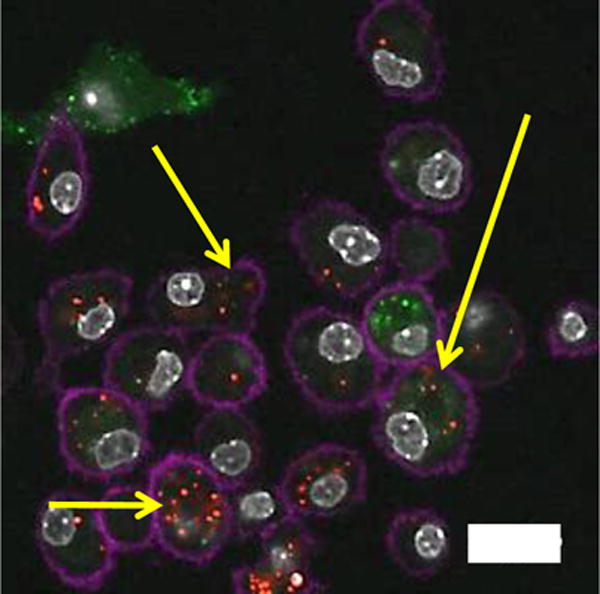
Laser scanning confocal microscopy imaging of cell-particle hybrid uptake by BMDC. Cell-particle hybrids were incubated with BMDC Yellow arrows indicating colocalization of B16.F10 cells (green) and particles (red) inside BMDC (magenta). Magenta: Alexa flour®700 (CD11c) stained BMDC, green: CFSE labeled B16.F10 melanoma cells, red: rhodamine B-labeled PLGA particles, gray: DAPI stained cell nuclei. Scale bar: 20 micron.