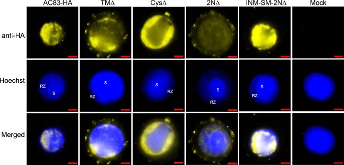
FIG 9.
Localization of AC83 from AC83HA, TMΔ, CysΔ, 2NΔ, and INM-SM+2NΔ in T. ni BTI-Tn5B1-4 cells. BTI-Tn5B1-4 cells were infected at an MOI of 5 and fixed at 48 hpi. Fluorescence microscopy was used to examine AC83 localization (yellow), and the nucleus was stained with Hoechst 33342 (blue). Noninfected cells were used as a control (Mock). Arrows indicate AC83 foci at the plasma membrane. RZ, ring zone; S, virogenic stroma. Cells were immunostained with rabbit anti-HA monoclonal antibodies followed by goat anti-rabbit IgG antibodies conjugated with Alexa Fluor 680 (yellow). The scale bar is 5 μm.

