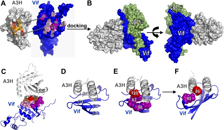
FIG 6.
Identification of the Vif-A3H interface by structural docking. (A) Overview of A3H and Vif residues important for the Vif-A3H interaction. Vif and A3H were docked using ClusPro. (B) The structure that has A3H positions 86 and 129 in close proximity to Vif positions 45 and 63 to 90 is shown. CBF-β is indicated in green and does not overlap A3H. (C) A cartoon model of the Vif-A3H structure in which the important Vif and A3H are indicated shows that they all localize to the Vif-A3H interface. (D) Close-up of the Vif-A3H interface. A3H α-helixes α3 and α4 interact with the Vif β-sheet consisting of β-strands β2 to β5. The model indicates that Vif-45 and A3H-86 (E) and Vif-63/90 and A3H-129 (F) are in close proximity.

