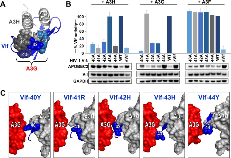
FIG 8.
Validation that Vif β-strand 2 is important for interaction with A3H and A3G. (A) Close-up of the A3H α-helixes α3 and α4 interacting with Vif residues 40 to 44. Residue colors correspond with the graph indicated in panel B. (B) Single-cycle infectivity assays of the indicated Vif mutants together with 40 ng A3H, A3G, or A3F. The infectivity was analyzed by TZM-bl reporter cells. Infectivity values are relative to the infectivity of WT HIV in the presence of A3H/A3G/A3F, which was set at 100%. The average relative infectivity values for a triplicate transfection are shown, and error bars represent the standard deviations. Cell lysates were analyzed by Western blotting. (C) The Vif-A3H structure is shown together with the N-terminal domain of A3G as described in Letko et al. (17). The individual Vif amino acids at positions 40 to 44 are indicated in blue together with A3H and A3G.

