
FIG 1.
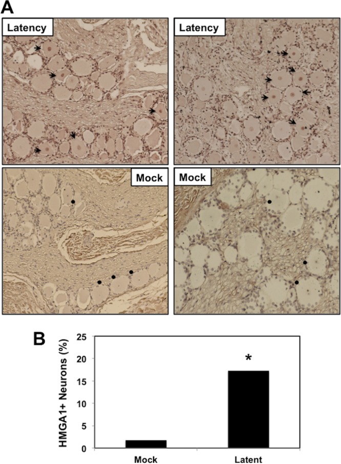
Detection of HMGA1 in TG neurons during latency. (A) TG were collected from 3 uninfected calves (denoted as Mock) or latently infected calves (at least 60 days postinfection). Thin sections were cut from formalin-fixed paraffin-embedded TG sections. The HMGA1 antibody used for this study was purchased from Abcam (catalog number ab129153) and was diluted 1:400. Biotinylated goat anti-rabbit IgG (Vector Laboratories) was used as a secondary antibody. Arrows, HMGA1-positive neurons; circles, TG neurons in uninfected calves that contain a visible nucleus in which the nucleolus was counterstained (however, the HMGA1 antibody did readily stain the nucleus). (B) The percentage of HMGA1-positive neurons among 490 total neurons was estimated from sections derived from three latently infected calves or three uninfected calves. *, significant differences (P < 0.05) in the numbers of HMGA1-positive neurons, as determined by a Student t test.