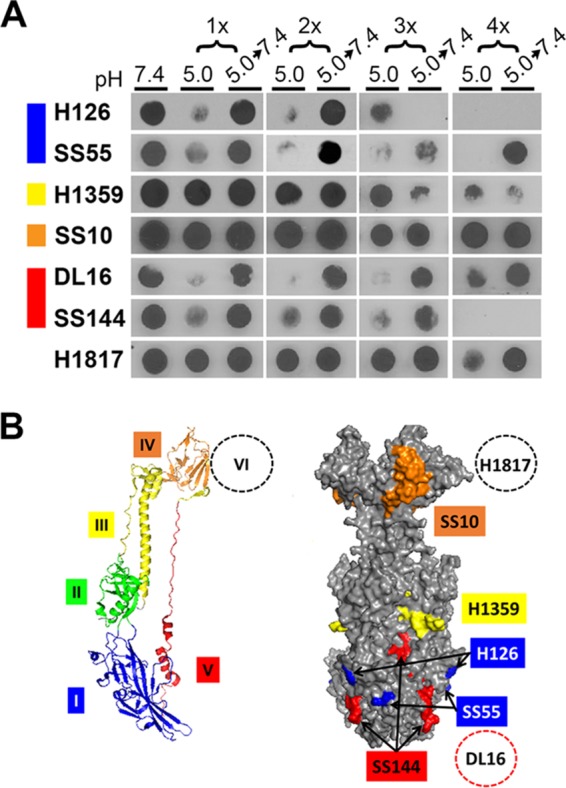
FIG 6.

Conformational change in gB from virions that have undergone a series of low-pH treatment reneutralization cycles. (A) Extracellular HSV-1 KOS virions (106 PFU) were treated with pH 5.0 for 10 min and neutralized back to pH 7.4 for 10 min for the indicated number of times. Samples were immediately blotted to nitrocellulose and probed at neutral pH with the indicated monoclonal antibody to gB. Each blot shown is representative of at least three independent experiments. (B) gB monomer with specific domains indicated by color (left) and space-filling model of gB trimer with surface-exposed epitopes detected by gB-specific monoclonal antibodies (right) (42). Domain I MAb H126 maps to residue 303 (42), and MAb SS55 maps to residues 203, 335, and 199 (51). Domain III MAb H1359 maps to residues 457 to 475 (49). Domain IV MAb SS10 maps to residues 640 to 670 (50). Domain V MAb SS144 maps to residues 697 to 725 (42), and MAb DL16 maps to residues 678 to 730 (51). Domain VI MAb H1817 maps to residues 31 to 43, which are unresolved in the crystal structure (50).
