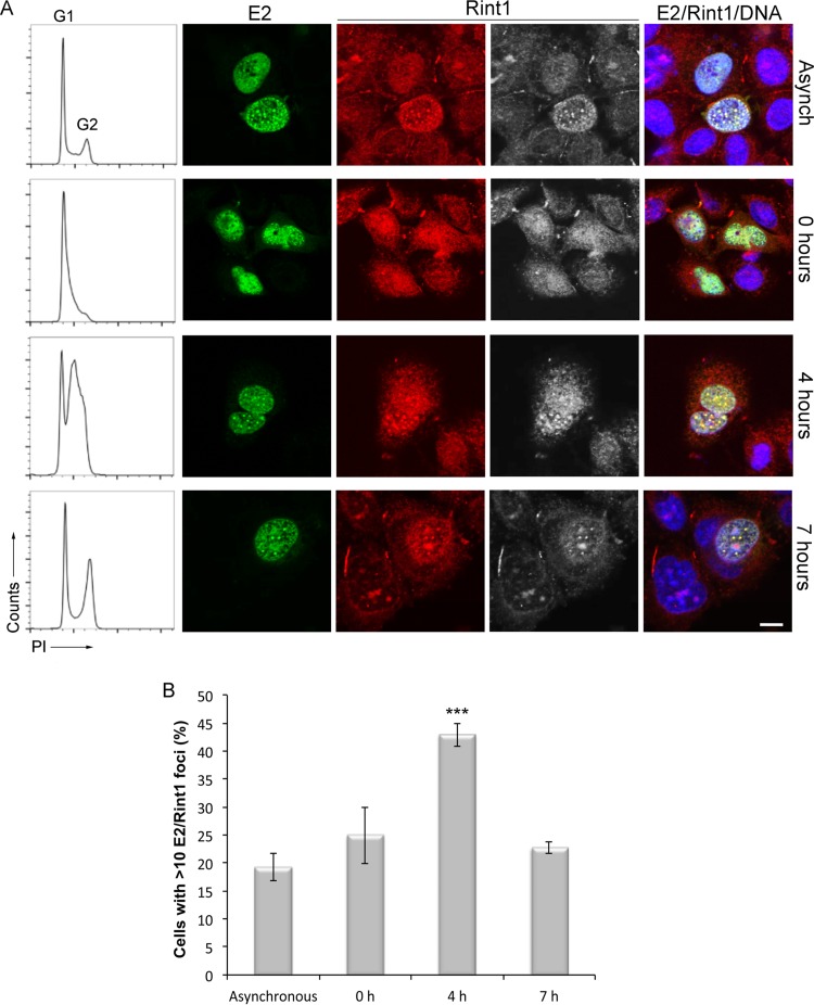
FIG 6.
HPV16 E2 and Rint1 colocalize in nuclear foci during S phase. C33a cells were transfected with an HPV16 E2-expressing plasmid and synchronized by a double-thymidine block. Asynchronously growing cells or cells harvested 0, 4, or 7 h following release were fixed and stained with propidium iodide (PI), and cell cycle profiles were obtained by flow cytometry (left). Cells within the same cultures were fixed at the indicated time points. Localization of E2 and Rint1 was determined by staining with the E2-specific TVG261 antibody (green) and Rint1 antibody (red/gray). DNA was stained with Hoechst 33342 (blue). Bar, 5 μm. (B) Quantification of E2-expressing cells containing >10 E2/Rint1-positive foci at each time point. A total of 500 cells were scored at each time point. The data shown are the means and standard deviations from three independent experiments (***, P < 0.001 compared to asynchronous cells).

