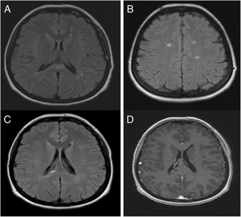
Fig. 1.

Brain MRI lesions of 52 year-old patient with CAPS before and after treatment. Axial FLAIR brain MRI images showing well limited periventricular (a), corpus callosum (a) and juxtacortical lesions (b). Brain MRI images with Axial FLAIR sequences (c) and T1 after gadolinium injection sequences (d), after one year of canakinumab treatment, of the same patient showing new FLAIR right paraventricular abnormalities with Gadolinium enhancement
