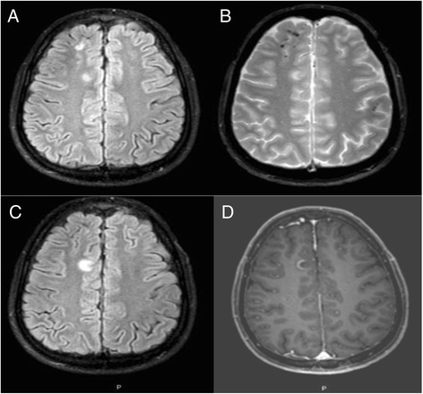
Fig. 2.

MRI images of 26 year-old patient with CAPS during a relapse with severe headaches. (a): Axial Flair images showing T2 FLAIR hypersignal right frontal juxta-cortical lesions with (b) hyposignal lesions on T2* weighted sequences suggesting hemosiderin deposits and (c) right anterior cingular gyrus T2 FLAIR hypersignal with (d) gadolinium enhancement
