
Figure 4. In vivo inactivation of PV neurons by DREADDs for 24 hours shows a large reduction in local excitatory input, and this is restored by bath NRG1 application.
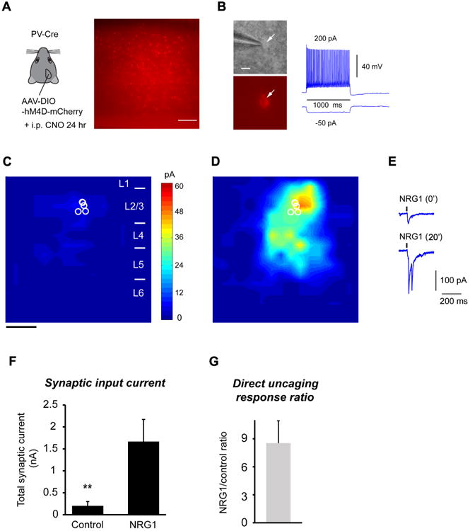
A, Use of the DREADDS to reduce PV neuron activity in vivo, phenocopies the effect evoked by monocular deprivation. Left, a schematic of injecting AAV-DIO-hM4D-mCherry in binocular V1 of the PV-Cre mouse. Right, a representative slice image showing AAV labels by mCherry visualization in PV neurons. Scale = 200 μm. B, Left: Targeted recordings of PV neurons are facilitated by mCherry expression in PV-Cre mouse slices. Right:In vivo CNO treatment does not affect the general fast-spiking phenotype of PV neurons in in vitro recordings. Scale = 10 μm. C-D, Group-averaged, excitatory input maps of L2/3 PV cells (n = 6 cells) are shown for before (C) and during bath NRG1 (D). The spatial scale beneath (C) indicates 200 μm. E, example responses evoked by photostimulation in the same perisomatic site before and during bath NRG1. F, Summary data of average total synaptic input strength measured for L2/3 PV neurons before (control) and during bath NRG1. **, p < 0.01 (Mann–Whitney U test). G, Direct uncaging response ratio of during bath NRG1 for DREADDs-inhibited PV cells versus before NRG1 application.