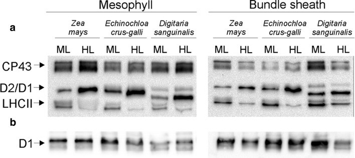
Fig. 5.
PSII protein phosphorylation in mesophyll and bundle sheath thylakoids isolated from the leaves of Zea mays, Digitaria sanguinalis, and Echinochloa crus-galli. The leaf samples were collected 2 h after growth light (ML) was turned on, and after that leaves were shifted for 1 h to high light (HL, 1600 μmol photons m−2 s−1) from ML (a). Gel wells were loaded with 1.5 µg of Chl equivalents of the isolated thylakoids. Phosphorylated proteins were detected with anti-PThr antibody. The positions of detected phosphoproteins are indicated on the left side. The level of D1 protein in thylakoids was confirmed using D1 protein-specific antibody (Agrisera) (b). Thylakoid proteins (1.0 µg Chl) were separated by urea SDS-PAGE and immunodetected. The results shown are representative of those obtained in at least three independent experiments. Thylakoids were isolated in the presence of 10 mM NaF

