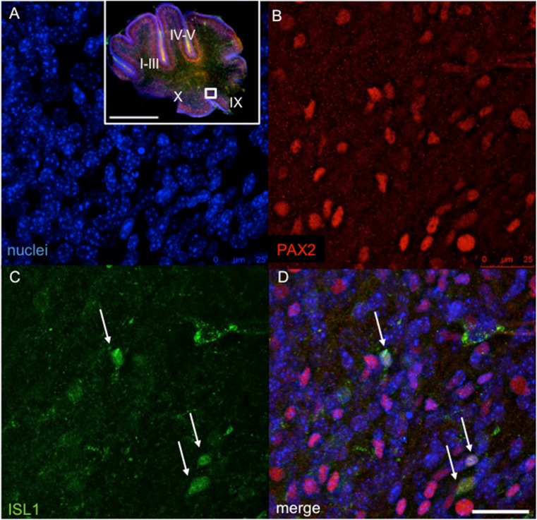
Fig. 10.
The expression of Isl1 in the transgenic cerebellum at P3. Confocal microscopy of 100 μm sections shows the expression of Isl1 in the transgenic cerebellum (lobule IX) indicated by white arrows. Double staining with anti-Pax2 (b, red) and anti-Isl1 (c, green) and visualization of nucleus with Hoechst staining (a) and overlay of fluorescent channels (d). Scale bar 500 μm (whole cerebellum), 25 μm (detail a–d)

