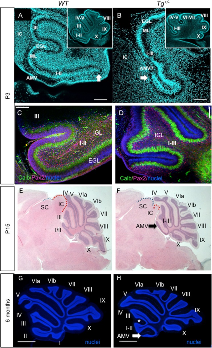
Fig. 4.
Changes in the cerebellum. a, b P3 sagittal sections using Hoechst nuclear staining show the different organizations in the control (WT) and mutant (Tg +/−) littermate cerebellar foliation (insert a, b) and disorganization of lobule I + II. Note absence of a recognizable anterior medullary velum (AMV) and the rostral expansion of a hemilobe only in the transgenic mouse (arrowhead). c, d Pax2 (red) and calbindin (green; Purkinje cells) staining of sagittal sections of the anterior lobe of the cerebellar vermis at P3 shows a comparable distribution of Purkinje cells and Pax2+ cells in WT (C) and Tg +/− lobules (d). The altered foliation of lobules I–III is obvious in the Tg +/− cerebellum. e, f Hematoxylin-eosin staining of the brain sections at the level of vermis at P15. The predominant phenotype of altered formation of vermis lobules leading to the fusion of I–III and a hemilobule on top of or as part of the anterior medullary velum (arrow) is detected in the Tg +/− cerebellum. The remnant of the inferior colliculus (IC) is denoted by a red asterisk in the Tg +/− midbrain. The superior colliculus (SC) and IC are outlined by blue- and red-dashed lines, respectively. g, h The adult Tg +/− cerebellum shows the defect in the foliation of the anterior lobe compared to WT littermates as shown by Hoechst staining of the granule cell layer nuclei. The fissure (*) between anterior folia I/II and III failed to form properly, leading to the fusion of the lobules. A hemilobule is on top of or as part of the anterior medullary velum (arrow). The lobules IV–V in Tg +/− differ from controls. Roman numerals depict cerebellum lobules. AMV, anterior medullary velum; Calb, calbindin; EGL, external granule layer; IGL, internal granule layer; IC, inferior colliculus; ML, molecular layer; SC, superior colliculus. Scale bar 100 μm (a–d) and 1000 μm (e–h)

