Fig. 7.
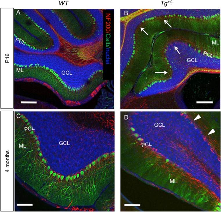
Changes in Purkinje cells in the anterior lobe (detail of lobules I–II). a, b Purkinje cells (PCs) are oriented in a monolayer with dendrites projecting into the ML at P16, as visualized by calbindin staining (green; nuclear staining, blue). More calbindin-negative PCs are visible in the Tg +/− anterior lobe (b, arrows). The density of PC dendrites stained by calbindin is noticeably reduced in Tg +/− compared to WT (a) at P16. c, d A profound reduction of calbindin expression in PCs and PC dendrites in the ML progresses with increasing age in the Tg +/− anterior lobe (d), as visualized by lack of staining with anti-calbindin. Anti-NF200 staining (red) of basket interneuron fibers wrapped around Purkinje cell bodies (arrowheads) is still detected in 4-month-old Tg +/− mice. ML, molecular layer; PCL, PC layer; GCL, granule cell layer. Scale bar 200 μm (a–b), 100 μm (c, d)

