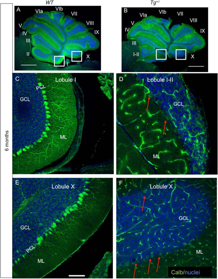
Fig. 8.
Reduction of Purkinje cell (PC) immunogenicity and apparent loss of PC dendrites in the molecular layer of the adult transgenic cerebella. PCs form a monolayer with dense network of dendrites in the ML throughout all the lobules in control cerebellum (a). At 6 months, a profound loss of calbindin expression in PCs and PC dendrites in the ML progresses in all lobules of the Tg +/− cerebella (b) as visualized by lack of staining with anti-calbindin (green). A near complete loss of calbindin expression in PCs and PC dendrites (arrows in d, f) is detected in the Tg +/− cerebella compared to WT, in detail shown in the lobules I–II and X (c, e). ML, molecular layer; PCL, PC layer; GCL, granule cell layer. Scale bar 1000 μm (a, b); 250 μm (c–f)

