Fig. 9.
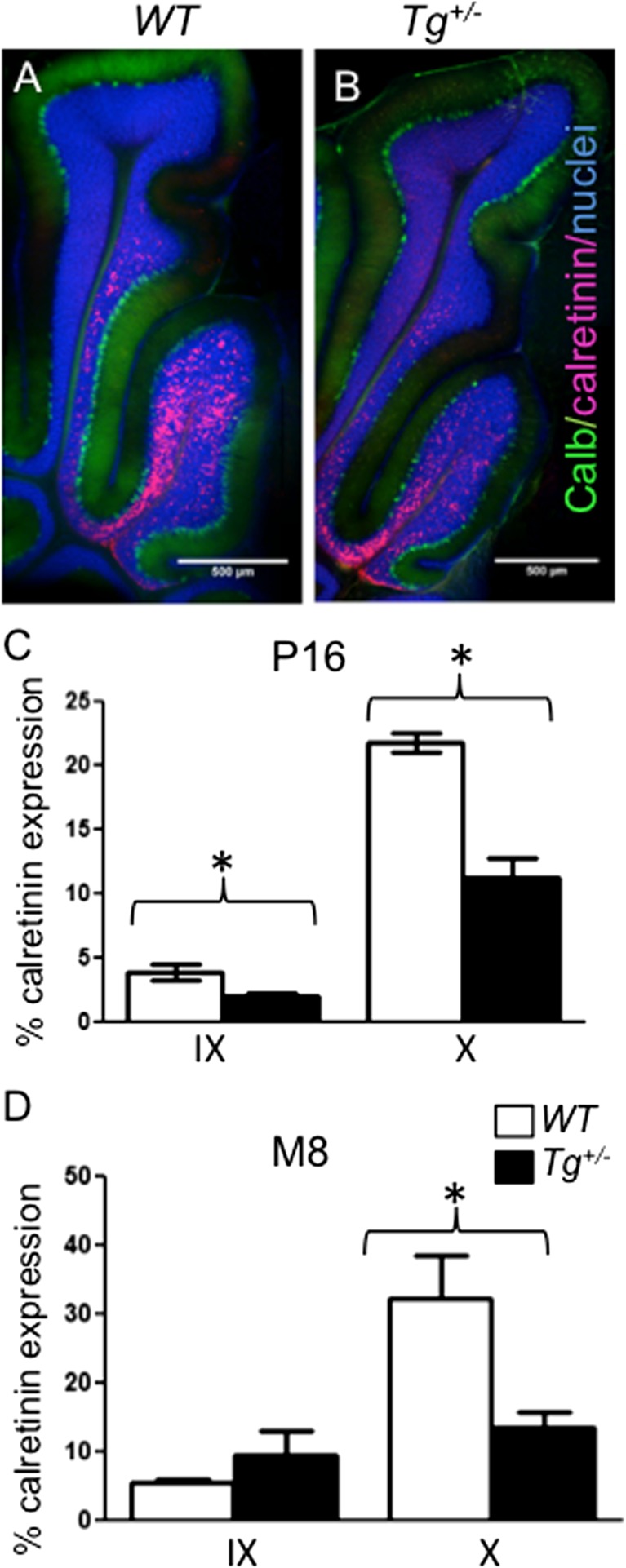
Altered distribution of calretinin-labelled cells in lobules X and IX of the transgenic cerebellum. Calretinin+ cells are primarily found in lobules X and half of IX as shown by calretinin staining (red) in both WT (a) and Tg +/− (b) cerebella. Double staining with anti-Calbindin (Calb, green) and anti-Calretinin (red) and visualization of nuclei with Hoechst staining of 100 μm sections of P16 cerebella. Scale bar 500 μm. Quantification of calretinin staining in lobules IX and X of the cerebellum at P16 (c) and 8-month-old (d) using ImageJ. The values represent an average percentage of calretinin+ area/lobule area ± SEM (n = 6 Tg +/− and 6 WT/each age group), t test *, P < 0.05

