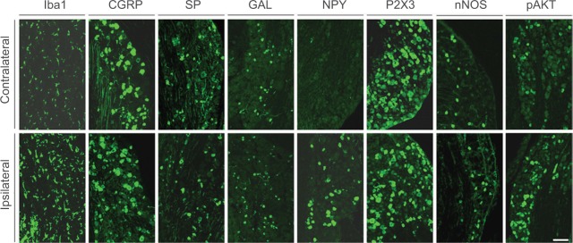
Figure 3.
Immunofluorescent micrographs show the expression of biomarkers in contralateral (top panel) and ipsilateral (bottom panel) TGs 2 weeks after partial ION transection.
Notes: Sections were incubated with antisera of Iba1, CGRP, SP, GAL, NPY, P2X3, nNOS, and pAKT, respectively. Scale bar indicates 100 μm.
Abbreviations: CGRP, calcitonin gene-related peptide; ION, infraorbital nerve; nNOS, neuronal nitric oxide synthase; NPY, neuropeptide Y; pAKT, phosphorylated AKT; SP, substance P; TGs, trigeminal ganglia.

