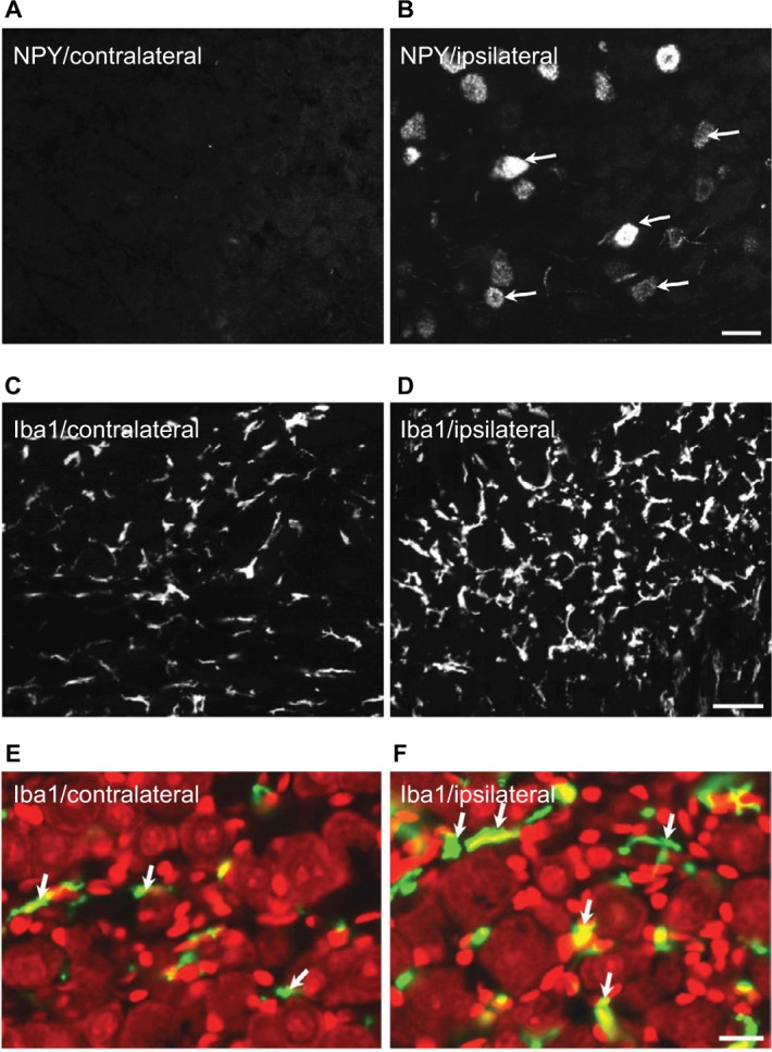
Figure 4.

Expression of NPY and Iba1 in ipsilateral and contralateral TGs 2 weeks after partial ION transection.
Notes: NPY-LI is upregulated in ipsilateral TG (B) compared with the contralateral one (A). Immunofluorescence micrographs show a hypertrophic morphology of Iba1-LI in ipsilateral (D and F) vs. contralateral TGs (C and E), counterstaining with PI (red) (E and F). Arrows indicate the NPY-positive neurons (B) and Iba1-positive microglial cells (E and F), respectively. Scale bars indicate 50 μm (A and B), 30 μm (C and D), and 25 μm (E and F).
Abbreviations: ION, infraorbital nerve; LI, like immunoreactivity; NPY, neuropeptide Y; PI, propidium iodide; TGs, trigeminal ganglia.
