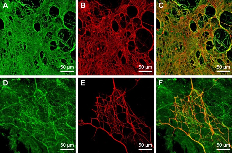
Figure 6.
Immunofluorescence staining of protein nanocoating on membrane.
Notes: Fibrin nanocoating (A), collagen nanocoating (D), fibronectin on fibrin nanocoating (B), fibronectin on collagen nanocoating (E), freshly deposited on poly(lactide-co-glycolic acid) membranes. Image A was merged with B (C), and image D with E (F). Secondary antibodies were conjugated with Alexa 488 (fibrin, collagen, green fluorescence) or with Alexa 633 (fibronectin, red fluorescence). Leica TCS SPE DM2500 confocal microscope, magnification 40×/1.15 NA oil.

