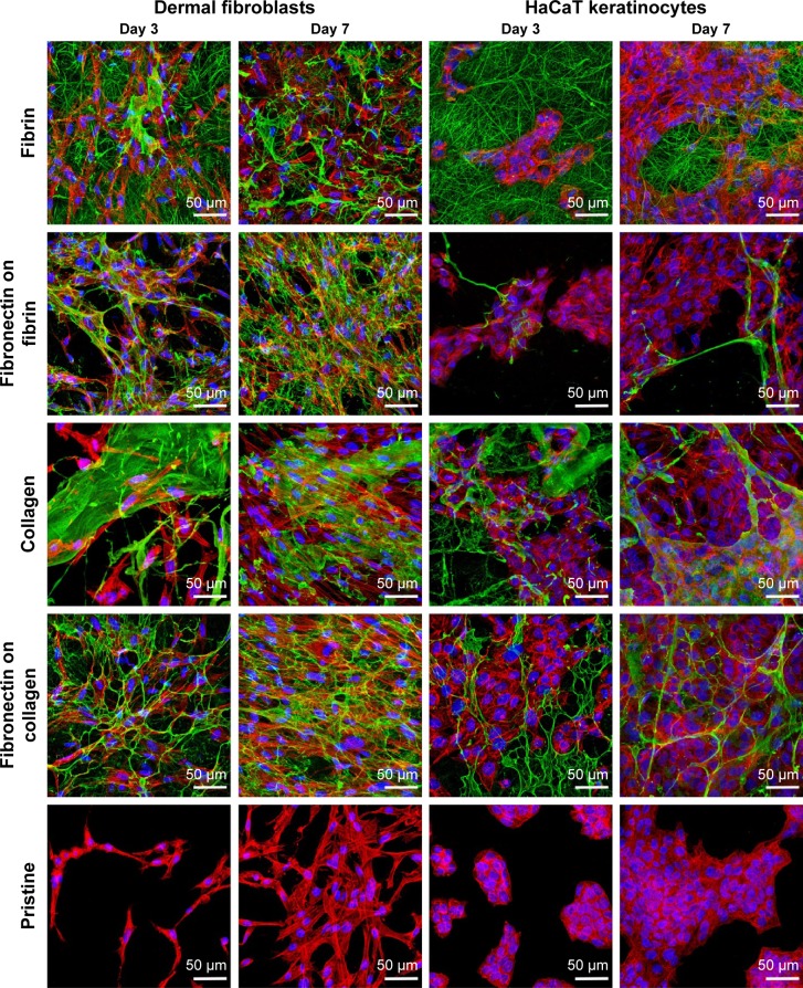
Figure 7.
Human dermal fibroblasts and HaCaT keratinocytes on protein-coated membranes.
Notes: Fibrin (row 1), fibronectin deposited on fibrin (row 2), collagen I (row 3), and fibronectin deposited on collagen (row 4) on poly(lactide-co-glycolic acid) membranes after 3 and 7 days of cultivation of human dermal fibroblasts and HaCaT keratinocytes. Row 5: control cells on pristine uncoated membranes. The protein nanocoating was immunofluorescence stained (green) with primary and secondary antibody conjugated with Alexa 488. The cells were stained with phalloidin–tetramethylrhodamine isothiocyanate (red; actin cytoskeleton) and with Hoechst 33258 (blue; cell nuclei). Leica TCS SPE DM2500 confocal microscope, magnification 40×/1.15 NA oil.

