Figure 6.
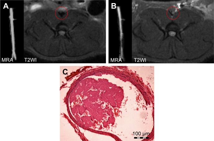
MRA, magnetic resonance imaging T2WI axial images, and pathologic H&E staining of the abdominal aorta.
Notes: (A) Before induction of the thrombosis model, (B) post-induction, and (C) H&E staining of a section of the model. MRA revealed vessel lumen narrowing after the FeCl3-induced model was successfully constructed, and the signal in the abdominal aorta changed from low to high on the T2WI axial image. H&E staining confirmed that the mural thrombus was mixed. The magnification is 100×.
Abbreviations: H&E, hematoxylin and eosin; MRA, magnetic resonance angiography; T2WI, T2-weighted imaging.

