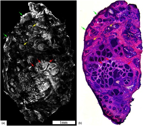
Fig. 1.
Acetic acid stained reflectance confocal mosaic (a) shows nodular BCCs (red arrows) that can be readily identified at magnification and compares well to (b) the corresponding H&E frozen histopathology. Nests of tumor (red arrows) within the dermis, the epidermal margin (green arrows) and hair follicles (yellow arrows) are seen. Scale bars: . Figure reproduced with permission, courtesy of Wiley.15

