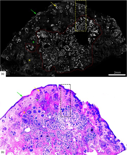
Fig. 4.
Acridine orange stained fluorescence confocal mosaic of a micronodular BCC (a) that equates well to view on standard H&E stained histology section (b). Small and tiny nodules or nests of tumor (red dotted area) can be identified at this magnification. Epidermis (green arrow), hair follicle (yellow arrow), sebaceous glands (green asterisk), dermal collagen (yellow asterisk) are seen. Scale bar: A = 2mm. Figure reproduced from Gareau et al. “Confocal mosaicing microscopy in Mohs skin excisions: feasibility of rapid surgical pathology.”14

