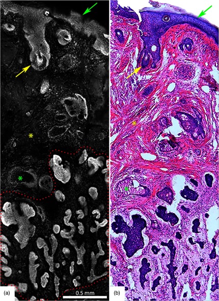
Fig. 5.
Acridine orange stained fluorescence confocal submosaic (a) obtained by digitally zooming ( magnification) in the mosaic (Fig. 4(a), dashed boxed area) to appreciate morphological features of micronodular BCC tumor (red dotted area) such as nuclear pleomorphism, increased nuclear density, and clefting. Epidermis (green arrow), hair follicle (yellow arrow), sebaceous glands (green asterisk), dermal collagen (yellow asterisk) are seen. This corresponds well with H&E stained frozen histopathology (b). A = 0.5 mm. Figure reproduced from Gareau et al. “Confocal mosaicing microscopy in Mohs skin excisions: feasibility of rapid surgical pathology.”14

