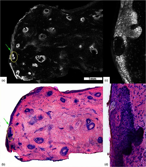
Fig. 6.
(a) Acridine orange stained fluorescence confocal submosaic shows a small bright focus (dotted circle) of superficial BCC along the epidermal margin (green arrow) raising suspicion for residual tumor and (b) corresponding H&E-stained frozen histopathology at magnification. Digital zooming to higher magnification () reveals the subtle but characteristic features of (c) superficial BCC on confocal image as well as on (d) H&E-stained frozen histopathology. This figure demonstrates that small foci of superficial BCC may be sometimes missed on confocal imaging either due to their subtle morphological features or uneven flattening of the tissue. Scale bar: . Figure reproduced with permission, courtesy of Wiley.34

