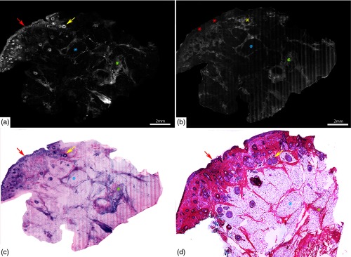
Fig. 8.
DSCMs of normal skin tissue closely mimicking H&E stained tissue histology. Mosaic of normal skin tissue in (a) fluorescence mode, (b) in reflectance mode, and (c) digital H&E (DHE) image created by overlapping fluorescence and reflectance channels. Nucleated structures, such as epidermis (red arrows) and hair follicle (yellow arrow), appear bright in fluorescence mode (a) and dull-grey on reflectance mode (b). Adipocytes (blue asterixis) appear darker and well defined in the fluorescence mode than in reflectance mode. By contrast, dermal collagen (green asterixis) appears bright in both the modes. (d) The H&E image () compares well with the DHE image (c). Scale bars: A, .

