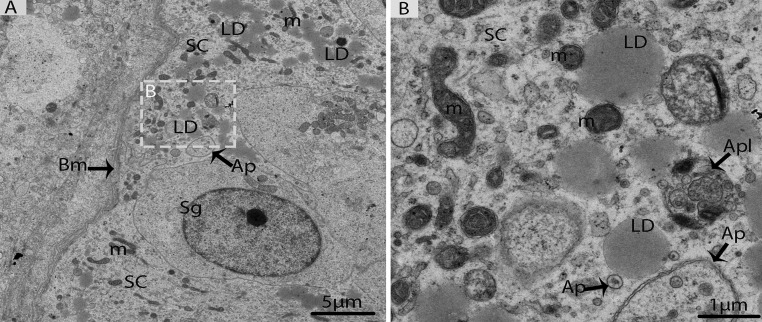
Figure 8. Electron micrograph of Sertoli cells in October.
(A) Sertoli cells appeared with lipid droplets mitochondria and autophagosomes. (B) Illustration of panel A (rectangular area) clearly shows the mitochondria and autophagosomes attached to lipid droplets. SC: Sertoli cell; Sg: spermatogonia; Bm: basal membrane; Ap: autophagosome; Apl: autophagolysosome; LD: Lipid droplets; m: mitochondria. Scale bar= 5μm (A) and1μm (B).

