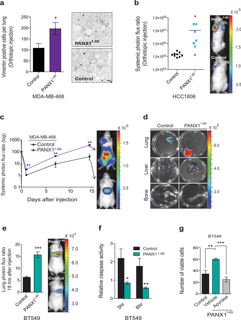
Figure 5. Mutational augmentation of PANX1 channel activity enhances the metastatic efficiency of breast cancer cells.
a, The numbers of vimentin positive breast cancer cells in the lung were counted one week after the extraction of size-matched mammary fat pad primary tumours generated by the orthotopic injection of 2.5× 10⁵ MDA-MB-468 cells expressing PANX1 1–89 or control vector into NOD scid gamma (NSG) mice; n = 4. Scale bar, 100 µm. b, Quantitative bioluminescence imaging of systemic metastasis one week after the extraction of size-matched mammary fat pad tumours generated by the orthotopic injection of 5 × 10⁵ HCC1806 breast cancer cells expressing PANX1 1–89 (n = 7) or control vector (n = 9) into NSG mice. c, Quantitative bioluminescence imaging of systemic metastasis after tail-vein injection of 1 × 10⁶ MDA-MB-468 breast cancer cells, expressing PANX1 1–89 or control vector, into NSG mice; n = 4. d, Ex vivo bioluminescence imaging of metastatic target organs (lung, liver and bone) 14 days after tain-vein injection of MDA-MB-468 cells. e, Quantitative imaging of lung bioluminescence 18 hrs post tail-vein injection of 1 × 10⁶ BT549 cells, expressing PANX1 1–89 or a control vector, into NSG mice; n = 6. f, In vivo quantification of luciferase-based caspase-3/7 activity at 3 and 6 hrs post tail-vein injection of 1 × 10⁶ BT549 cells, expressing either PANX1 1–89 or a control vector, into NS mice; n = 4 (3hr control), n = 5 (3hr PANX1 1–89), n = 4 (6hr control), n = 5 (6hr PANX1 1–89). g, Quantification of viable, trypan blue-negative BT549 cells expressing PANX1 1–89 or a control vector after 1 hr extreme hypotonic (12.5% PBS) stretch in the presence of succinate buffer or apyrase (2U/ml); n = 3. Error bars, s.e.m., *, P < 0.05; **, P < 0.01; ***, P < 0.001 by a one-tailed Student’s t-test. n represents biological replicates. Orthotopic experiments replicated in two independent triple-negative cell human breast cancer cell lines. Experiments e–g are representative and were replicated at least two times in at least two independent cell lines. Bioluminescent and histological images are representative of the median.

