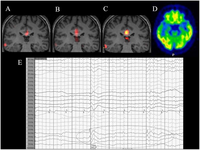
Fig 2. A 38-year-old man diagnosed with right lateral temporal lobe epilepsy.
(A) Coronal fALFF, (B) ALFF, and (C) ReHo reveal abnormal activation in the right lateral temporal lobe. An area of activation could also be found in the corpus callosum, which is considered a part of the default mode network. (D) The corresponding 18F-FDG PET/CT image reveals low FDG uptake in the right lateral temporal lobe region. (E) VEEG depiction of interictal epileptiform discharges of right temporal lobe.

