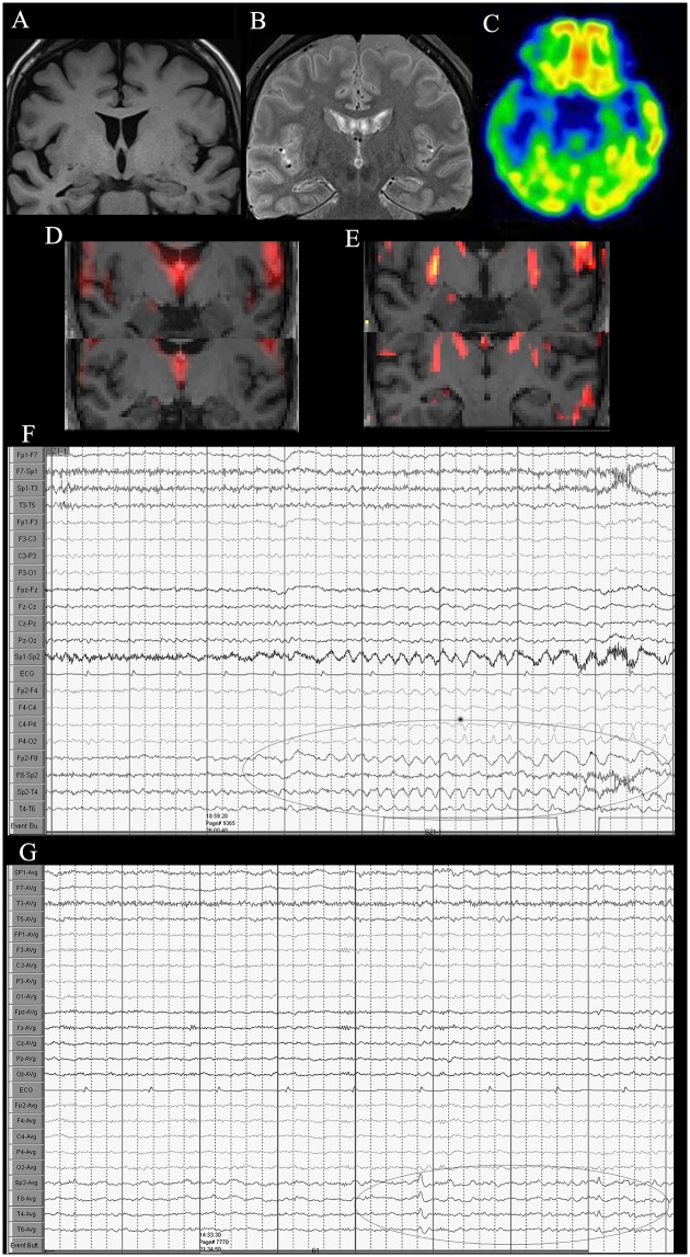
Fig 3. A 42-year-old woman diagnosed with right mesial temporal lobe epilepsy.
(A) Coronal T1-weighted image showing right hippocampus atrophy, (B) FLAIR image showing slightly high signal of right hippocampus. (C) 18F-FDG PET/CT imaging reveals low FDG uptake in the left temporal lobe region, particularly, in the hippocampus. (D) Coronal ALFF and (E) ReHo reveal abnormal activation in the right hippocampus compared to the left side. (F, G) VEEG showing ictal and interictal discharges in the right temporal lobe.

