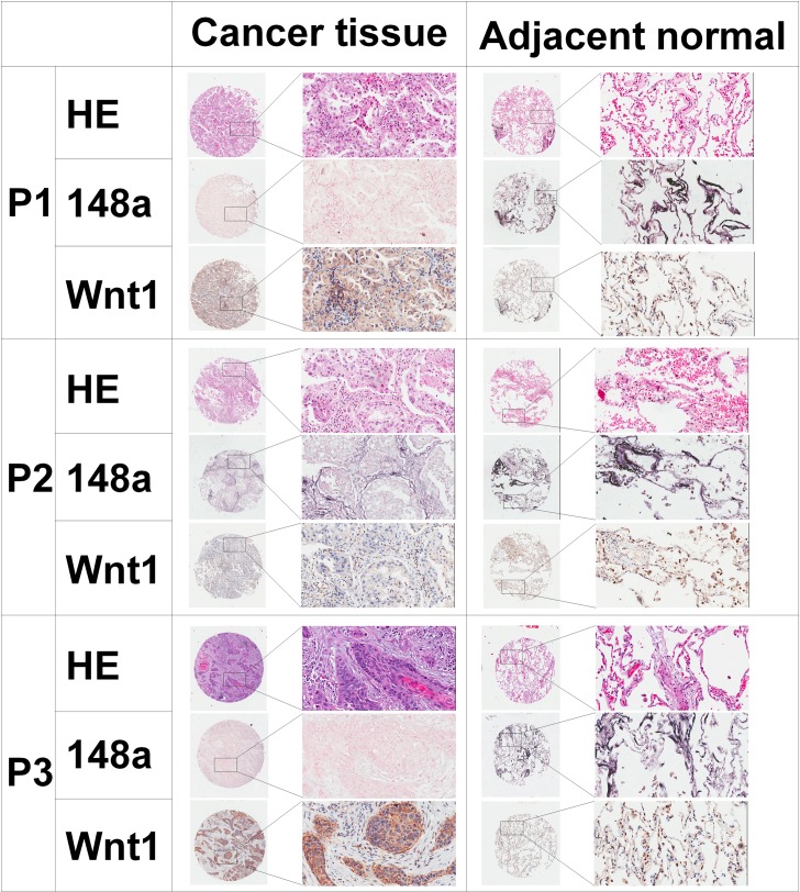
Fig 1. HE, ISH and IHC analysis of NSCLC tissue and corresponding adjacent normal lung tissues from 3 representative patients (P1 and P2: adenocarcinoma; P3: squamous cell carcinoma).
HE, ISH for miRNA-148a and IHC for Wnt1 from the same tissue area are shown. ISH shows differential expressions of miRNA-148a in primary tissues compared to adjacent normal lung tissues. Weak miRNA-148a staining in cancer tissues with corresponding strong miRNA-148a staining in adjacent normal lung tissues (P1 and P3). Negative correlation between miRNA-148a and Wnt1 expression in NSCLC samples. Increased Wnt1 immunoreactivity in cancer tissues with low levels of miRNA-148a assessed by ISH (P1 and P3). Decreased Wnt1 immunoreactivity in lung cancer tissues with high level of miRNA-148a (P2). Increased miRNA-148a staining in adjacent normal lung tissues with low levels of Wnt1 staining (P1, P2 and P3). HE: hematoxylin-eosin staining, ISH: in situ hybridization and IHC: immunohistochemistry.

