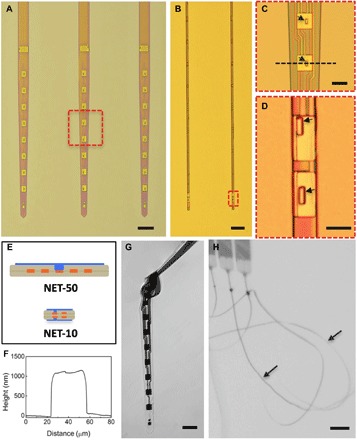
Fig. 1. Structures of NET neural probes.

(A and B) As-fabricated NET-50 and NET-10 probes on substrates. (C and D) Zoom-in views of two electrodes as marked by the dashed boxes in (A) and (B), respectively. Arrows denote “vias.” (E) Schematics of the probe cross section in (A, top) and (B, bottom), highlighting the multilayer layout. Color code: gray, insulation; orange, interconnects; and blue, electrodes. Not drawn to scale. (F) Height profile of the NET-50 probe along the dashed line in (C) measured by an atomic force microscope. (G) A NET-50 probe suspended in water. A knot is made with a curvature of less than 50 μm to illustrate its flexibility and robustness. (H) Multiple NET-10 probes suspended in water. Arrows denote the probes. Scale bars, 100 μm (A), 50 μm (B, G, and H), and 10 μm (C and D).
