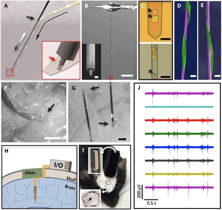
Fig. 2. Implantation procedure for the NET probes.

(A) Schematic showing the temporary engaging mechanism. Arrows denote the entry site of the implantation (solid), the delivery path of the shuttle device (gray), and the path of the engaged NET probe (dashed). Inset: Zoom-in view of the dashed square highlighting that the micropost engages in the microhole on the NET probe at the end of the shuttle device. (B) Photograph of a typical carbon fiber shuttle device, with a diameter of 7 μm and a length of 3 mm, mounted at the end of a micromanipulator. Scale bar, 500 μm. Inset: Scanning electron microscopy (SEM) image of the micromilled post with a diameter of 2 μm and a height of 5 μm at the shuttle device tip. Scale bar, 2 μm. (C) Optical micrographs showing engaging holes in NET-50 (top) and NET-10 (bottom) probes with a slightly larger diameter than the post, as denoted by the arrows. (D and E) False-colored SEM images of NET-50 and NET-10 probes (green) attached on shuttle devices with a 20-μm tungsten microwire (D, purple) and a 10-μm carbon fiber (E, purple) showing their ultrasmall dimensions. Scale bars, 50 μm (D) and 20 μm (E). (F and G) Micrographs showing that both NET-50 and NET-10 probes were successfully delivered into the living mouse brain with minimal acute tissue damage. Arrows denote the delivery entry sites. Scale bars, 100 μm (F) and 50 μm (G). (H) Schematic of skull fixation that accommodates both connectors for the neural probes and a glass window allowing optical access. Not drawn to scale. (I) Photograph of a typical postsurgery mouse with implanted NET probes and a glass window mounted on top. Insets: top, image of a cable connector mounted on the skull; bottom, zoom-in view of the glass window in which the arrow denotes an implanted probe. (J) Typical unit activities recorded by eight electrodes on an implanted NET-50 probe. A high-pass filter (300 Hz) was applied.
