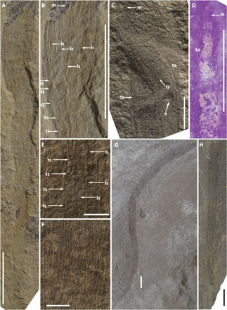
Fig. 5. Specimens and characteristic features of leptomitid protomonaxonid sponges from the Paris Biota.

(A and B) General and closeup view of the specimen UBGD 30504 showing projected longitudinal spicules (ls) from the apex forming a fringe of marginalia (m) and transverse spicules (ts). (C and D) Closeup views of twisted apex (ta) of two specimens (UBGD 30505 and 30581) under natural and UV light (365 nm). Projecting spicules from the apex forming a fringe of marginalia are also visible. Fine transverse spicules appear mainly as wrinkles, perpendicular to the longitudinal spicules. An epizoan brachiopod (e) is attached to the sponge specimen C. (E and F) Closeup views of specimens UBGD 30506 and 30508, showing longitudinal and transverse spicules. (G and H) Large-sized specimens UBGD 30510 and 30511. Scale bars, 5 mm (A to D and G and H), 2 mm (E), and 1 mm (I). [Photo credits: A. Brayard, Université Bourgogne Franche-Comté.]
