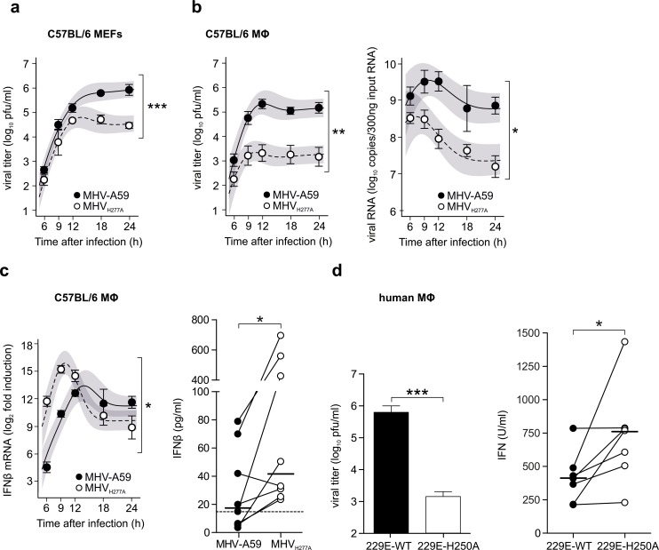
Fig 2. EndoU-deficient coronaviruses are severely attenuated in primary macrophages and trigger an elevated IFN-I response.
(a) Replication kinetics of MHV-A59 and MHVH277A in C57BL/6 mouse embryonic fibroblasts (MEFs) after infection at a MOI of 1, presented as viral titer in pfu. Data represent four independent experiments, each performed in two to four replicas. The difference in peak levels of viral titers (peak MHV-A59: 5.9, peak MHVH277A: 4.8) was statistically significant (***, p<0.001). (b) Replication kinetics of MHV-A59 and MHVH277A (left panel; titers in pfu) and cell-associated viral RNA (right panel; qRT-PCR) following infection of C57BL/6 bone marrow-derived macrophages (MOI = 1). Data represent eight (left panel) or five (right panel) independent experiments, each performed in two to three replicas. The difference in peak levels of viral titers (left panel: peak MHV-A59: 5.3, MHVH277A: 3.3) and the difference in peak levels of RNA copies (right panel: peak MHV-A59: 9.5, MHVH277A: 8.5) were statistically significant (p = 0.002, p = 0.018, respectively). (c) Expression of IFN-β mRNA (left panel; qRT-PCR) and protein (right panel; ELISA) in C57BL/6 macrophages following infection of MHV-A59 and MHVH277A (MOI = 1). Expression of IFN-β mRNA was normalized to levels of the household genes GAPDH and Tbp and is displayed relative to mock as ΔΔCT. The IFN-β ELISA detection limit is indicated with a dashed line. Data represent seven (left panel) or eight (right panel) independent experiments, each performed in two to three replicas. The difference in peak levels of IFN-β mRNA (MHV-A59: 13.4, MHVH277A: 15.8) was statistically significant (p = 0.04). Significance of IFN-β protein at 9 h.p.i. was assessed by a two-sided, Wilcoxon matched-pairs test (p = 0.016). (a-c) Mean and SEM are depicted. The 95% confidence band is highlighted in grey. Statistically significant comparisons are displayed; * p < 0.05, ** < 0.01, *** < 0.001. (d) Titers (pfu) of HCoV-229E wild type and HCoV-229EH250A (left panel) and expression of IFN-I (right panel; IFN-I bioassay) in human blood-derived macrophages, 24 hours after infection (MOI = 1). Data represent six (left panel) or seven (right panel) independent experiments, each performed in three to four replicas. Significance was assessed by a two-sided, unpaired Student’s t-test (left panel, p<0.001) and a Wilcoxon matched-pairs test (right panel; p = 0.016). (c-d) Mean and SEM are depicted. Statistically significant comparisons are displayed; * p < 0.05, *** < 0.001.

