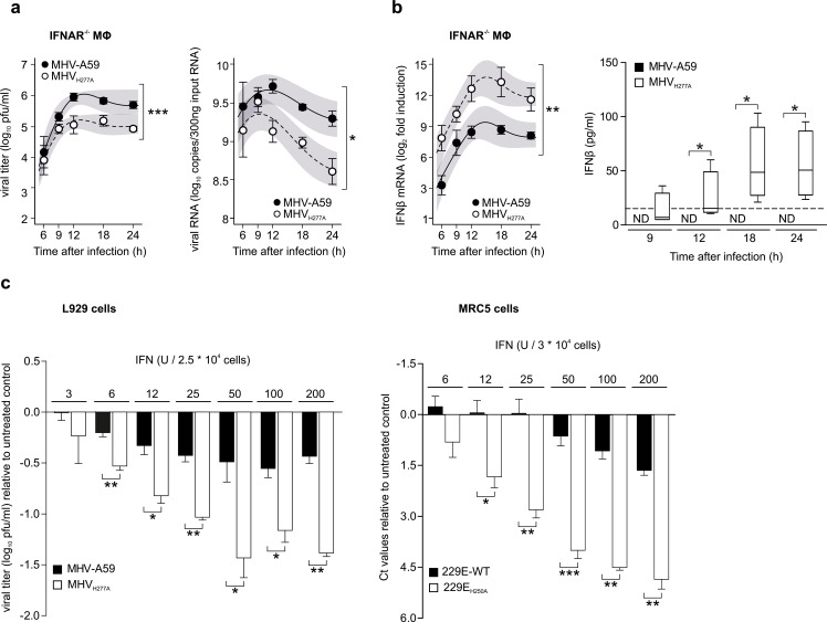
Fig 4. Replication of EndoU-deficient MHV is partially restored in IFNAR-/- macrophages and EndoU mutants display a pronounced sensitivity to IFN-I treatment.
(a) Replication kinetics of MHV-A59 and MHVH277A (left panel; titers in pfu) and cell-associated viral RNA (right panel; qRT-PCR) following infection of IFNAR-/- bone marrow-derived macrophages (MOI = 1). Data represent four independent experiments, each performed in two to three replicas. Mean and SEM are depicted. The 95% confidence band is highlighted in grey. The differences in peak levels of viral titers (MHV-A59: 6.0, MHVH277A: 5.2) and RNA copies (MHV-A59: 9.7, MHVH277A: 9.3) were statistically significant (p<0.001, p = 0.032, respectively). (b) Expression of IFN-β mRNA (left panel; qRT-PCR) and protein (right panel; ELISA) in IFNAR-/- macrophages following infection of MHV-A59 and MHVH277A (MOI = 1). Data represent four (left panel) and three (right panel) independent experiments, each performed in two to three replicas. Median and the 1–99 percentiles are displayed. Dashed line depicts limit of detection (right panel). The difference in peak levels of IFN-β expression (MHV-A59: 9.4, MHVH277A: 13.8) was statistically significant (p = 0.002). Significance of IFN-β expression was assesses by a Wilcoxon matched-pairs test, * p < 0.05. ND, not detected. (c) Sensitivity of wild type and EndoU-deficient MHV (left panel) and HCoV-229E (right panel) viruses to IFN-I pre-treatment (4 h) in L929 cells (left panel) and MRC-5 cells (right panel) with various dosages of IFN-I (MOI = 1). Virus replication was measured at 24 h.p.i. by plaque assay (MHV) and at 48 h.p.i. by qRT-PCR (HCoV-229E), respectively. Data represent three independent experiments, each performed in two to three replicas. Data are displayed as differences to untreated controls and statistical comparisons between wild type and EndoU-deficient viruses were performed for each concentration. Mean and SEM are displayed. Data points that show significant differences in a two-sided, unpaired Student’s t-test are depicted. * p < 0.05, ** p < 0.01 and *** p < 0001.

