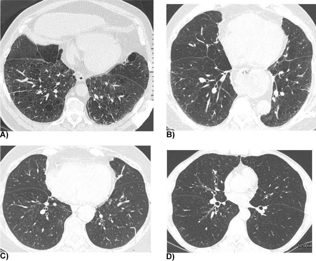
Fig. 2. Representative cases of the 4–point grading system for the visual assessment of airway disease.
(a) Grade 1 (absence of bronchial wall thickening). A 63-year-old man with a %LAA−856exp of 44.67%. (b) Grade 2 (borderline bronchial wall thickening). A 63-year-old woman with a %LAA−856exp of 46.56%. (c) Grade 3 (definitive bronchial wall thickening). A 74-year-old woman with a %LAA−856exp of 61.71%. (d) Grade 4 (severe bronchial wall thickening). A 56-year-old man with a %LAA−856exp of 65.65%.

