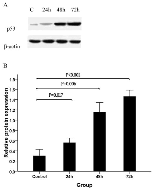
Fig. 6.
Effect of As2O3 on p53 protein expression in WSU-CLL cells. WSU-CLL cells were exposed to 2.0 μM As2O3 for 0, 24, 48, and 72 h. Western blot analyses were performed. (A) Representative immunoblots of the p53 expression level following treatment with 2 μM As2O3 for 0, 24, 48 and 72 h. (B) β-actin expression was used as a loading control. Relative p53 protein levels (means ± SD, n = 3) were determined.

