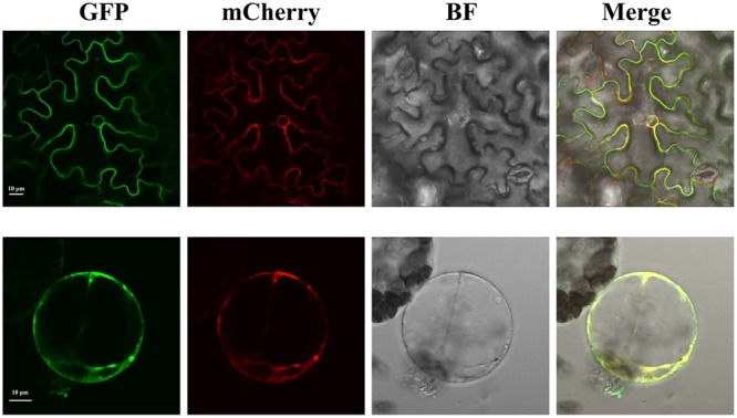
FIGURE 3.

Confocal images of the subcellular localization of SlTDT-GFP fusions in tobacco leaf mesophyll cell. The tonoplast marker TIP-RFP was used as the control. The GFP fusion protein is shown in green; the RFP fusion protein is shown in red. All images shown were acquired using the same photomultiplier gain and offset settings. Scale bar: 10 μm.
