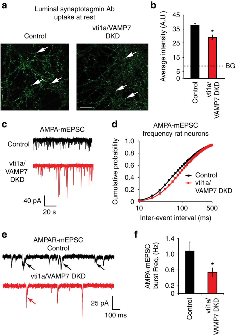
Figure 1. Vti1a and VAMP7 loss in cultured rat hippocampal neurons decreases synaptic vesicle trafficking at rest.
(a) Representative images of anti-luminal domain of synaptotagmin 1 antibody uptake at rest in control and vti1a/VAMP7 DKD (DKD) neurons. White arrows indicate representative areas selected for intensity analysis typical of presynaptic boutons. Scale bar, 10 μm. (b) Quantification of anti-luminal domain of synaptotagmin 1 antibody intensity at rest in control and vti1a/VAMP7 DKD neurons (control: n=167 puncta from 10 images from two independent cultures; vti1a/VAMP7 DKD: n=221 puncta from 11 images from two independent cultures; P=0.002). Background (BG; dashed line) fluorescence levels were obtained from similar experiments in which the primary antibody was omitted. (c) Representative traces of spontaneous AMPA event (AMPA-mEPSC) recordings in control and vti1a/VAMP7 DKD neurons. (d) Cumulative probability histograms of AMPA-mEPSC inter-event intervals (control: n=29 recordings from seven independent cultures; vti1a/VAMP7 DKD: n=31 recordings from seven independent cultures; Kolmogorov–Smirnov test; P=0.0001). (e) Representative traces of AMPA-mEPSC recordings containing mini-bursts (indicated by arrows) in control and vti1a/VAMP7 DKD neurons. (f) Average AMPA-mEPSC burst frequency was significantly decreased in vti1a/VAMP7 DKD neurons compared with control in the same recordings analysed in d (P=0.036).

