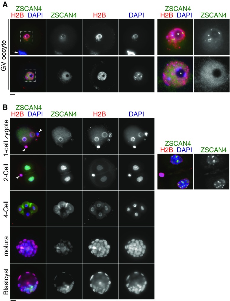
Figure 3.
Zscan4 protein is expressed in GV oocytes and preimplantation embryos. (A) GV oocytes were immunostained as indicated. Two types of immunostaining patterns in GV oocytes are shown. The upper example shows the spotty immunostaining pattern of Zscan4, some of which surround a nucleolus. The lower example shows the faint diffusive immunostaining pattern of Zscan4. Enlarged images of the nuclei are shown on the right. Arrow indicates cumulus granulosa cells associated with zona pellucida of GV oocyte. Asterisk indicates nucleolus. (B) Preimplantation embryos at different developmental stages were immunostained as indicated. Enlarged deconvolution images of partial Z projection for two cells are shown on the right, emphasizing intense Zscan4 foci. Arrowhead indicates polar body in two cells. Scale bars, 20 μm.

