Figure 4.

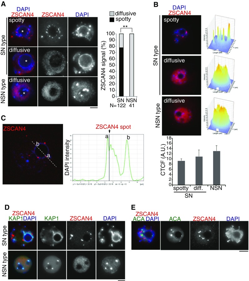
Zscan4 localization in SN- and NSN-type GV oocytes. (A) GV oocytes, classified as SN type and NSN type by heterochromatin morphology relative to nucleolus (shown by asterisk), were immunostained as indicated. The spatial localization pattern of Zscan4 was classified as spotty (limited number of foci) and diffusive (dispersed in nuclei). The immunostaining pattern of Zscan4 is quantified in the right graph. The number of analyzed GV oocytes (pooled from four independent experiments) is indicated. **P < 0.001 (Pearson’s chi-square test). (B) Intensity profile of Zscan4 immunostaining signal obtained with the FluoView Software from representative GV oocytes with a SN spotty (top, n = 21), SN diffuse (middle, n = 11), and NSN (bottom, n = 23). The value of pixel intensity on Z-axis is equal to 200 for the spotty signal (SN spotty) and to 180 (SN diffuse, NSN) for the oocytes with diffuse signal as inferable from the picture. Corrected total cell fluorescence (CTCF, arbitrary units) is shown in the bottom graph with SEM. C Zscan4 and DAPI signals were scanned across a nucleolus in SN-type GV oocyte (dashed line). Signal intensity of DAPI (green) and Zscan4 (gray bar) is shown on the bottom. D SN-type and NSN-type GV oocytes were immunostained for KAP1: a heterochromatic protein which is potentially associated with Zscan4. E SN-type GV oocyte was immunostained for anti-centromere antigen (ACA). Scale bars, 10 μm.
