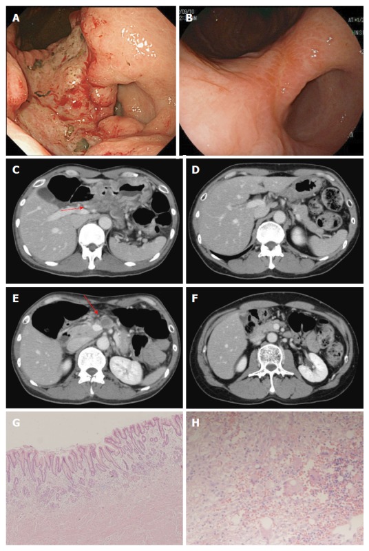
Figure 2.

Case 2. Endoscopic findings (A, B) and computed tomography images (C, D, E, F) before treatment (A, C, E) and after (B, D, F) five courses of modified docetaxel, cisplatin and capecitabine (DCX) (mDCX). The primary lesion (A, B), swollen lymph nodes along the common hepatic artery (#8) (C, D) and lymph nodes along the superior mesenteric artery (#14a) (E, F) markedly shrank. Microscopic findings for the resected specimens of the primary lesion (E, magnification × 100) and lymph nodes (F, magnification × 400) revealed no residual tumor.
